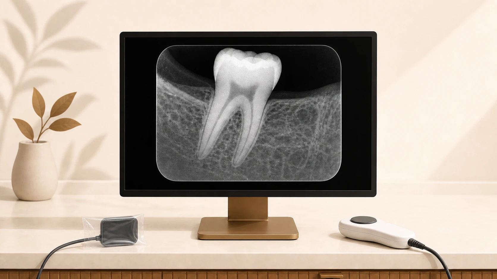
Viewing your X-ray on screen can help you understand what the dentist is assessing. Here is how the image is obtained, what it is used for, and its limits.
What is digital X-ray?
An electronic sensor receives X-rays passing through tissues and produces a digital image. The dentist can view it on screen, enlarge it, and adjust contrast. These tools assist interpretation but do not add information that is absent from the image.
Advantages over traditional film X-rays
- Exposure suited to the examination: it depends on the equipment and protocol. There is no identical percentage reduction for every comparison with film.
- Rapid image availability: no chemical processing is needed.
- Viewing tools: the image can be enlarged and its brightness and contrast adjusted for interpretation.
- Digital storage: your complete X-ray history stays in your electronic record, accessible for comparisons over time.
- No toxic chemicals: there are no developer or fixer liquids contaminating the clinic environment.
- Shareable: sent by email, WhatsApp for second opinions, consultations, or so you can see them yourself.
- Quality control: the clinician can promptly assess whether the image answers the clinical question or another view is needed.
What does your endodontist see in a dental X-ray?
Depending on image quality and projection angle, a periapical X-ray can help assess:
- Cavities between teeth (interproximal cavities) invisible to the naked eye.
- The shape and curvature of roots and canals visible in the image.
- Changes around the root tip that need to be interpreted alongside the examination.
- Internal or external root resorptions.
- Root fractures (in cases where the fracture displaces fragments).
- Previous root fillings and restorations, within the limits of two-dimensional imaging.
- Bone loss from periodontal disease.
- Remaining root fragments or areas requiring further assessment; an image alone does not establish that a lesion is a cyst.
- Impacted or supernumerary teeth.
- Bone levels near the tooth, without replacing three-dimensional assessment for implants.
Is it safe?
Dental X-rays use ionizing radiation. The clinician weighs their benefit, selects necessary views, and adjusts exposure. The ADA recommends selecting imaging according to clinical need.
- Bring previous X-rays so the clinician can assess whether they remain useful.
- Tell the team if you are pregnant or have questions about imaging.
- Ask which question the X-ray is intended to answer.
- The dose depends on the equipment, view, and protocol; it cannot be compared with a particular flight without those details.
When indicated, dental X-rays can be taken during pregnancy. Necessary imaging is selected and the applicable protocol followed.
When are X-rays taken in endodontics?
The number and timing of images depend on the case. They may be used for:
- Initial diagnostic X-ray: to view root anatomy and the problem requiring treatment.
- Assessing working length alongside the apex locator and clinical findings.
- Assessing the final root filling; the image alone cannot confirm a three-dimensional hermetic seal.
In cases of complex anatomy or accessory canals, additional X-rays may be taken to obtain different angulations.
Why does this matter specifically in endodontics?
Endodontics works inside the tooth, in millimetric and sometimes sub-millimetric spaces. A high-resolution digital X-ray allows the endodontist to:
- Reviewing visible anatomy before treatment.
- Looking for anatomical variations that may guide assessment.
- Supporting the determination of working length.
- Assessing treatment stages when imaging is needed.
- Document treatment for long-term follow-up.
X-rays are interpreted alongside the examination, microscope, and apex locator. If a CT scan or another study is needed, it is requested through referral; the clinic has digital two-dimensional periapical imaging.
What if I bring X-rays from elsewhere?
If you bring previous X-rays from another clinic or the United States, the clinician reviews whether they are useful and decides whether additional images are needed. They are not repeated automatically.
Technology in service of the patient
At Endodontics El Salvador in San Miguel, we explain evaluation findings and treatment options. You can ask what the image shows and what information needs to be confirmed by other tests.
Last updated: .